
FIGURE 3:
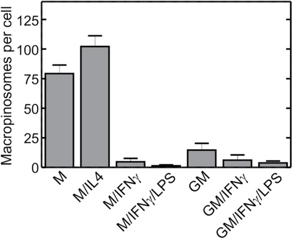
Macropinocytic activity of macrophages cultured under various conditions. Human blood monocytes were cultured for 5 d in either M-CSF (columns 1–4) or GM-CSF (columns 5–7), followed for 2 more days in either M-CSF (M) or GM-CSF (GM) alone, or in the additional presence of either IL-4 (M/IL4), IFN-γ (M/IFN-γ and GM/IFN-γ), or a combination of IFN-γ and LPS (M/IFN-γ/LPS and GM/IFN-γ/LPS), as described in Materials and Methods. On day 7 cells were incubated with fluorescently labeled 70 kDa dextran (TMR-dextran, 125 µg/ml) for 15 min at 37°C in calcium-containing medium. Cells were then washed, fixed, and imaged immediately by spinning disk confocal microscopy. The number of macropinosomes (i.e., TMR-positive vacuoles) per cell then was determined as described for Figure 2C. Data are means ± SEM of 30–75 cells from three to seven experiments of each type using blood from at least three different donors. Probabilities of statistical significance for comparisons between all pairs of data sets are presented as a matrix in Supplemental Table S2.