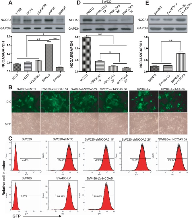
Figure 2. Lentivirus-mediated knockdown and overexpression of NCOA5 in CRC cells.
(A) Western blot analysis of NCOA5 in a panel of CRC cell lines. The lysates of HT29, HCT8, HCE8693, SW620 and SW480 CRC cells were immunoblotted with anti-NCOA5 or anti-GAPDH (a loading control) antibody. **, P<0.01 compared withHT29, HCT8, HCE8693 and SW480 group; *, P<0.05 compared withHT29, HCT8 and HCE8693 group, one-way repeated measures ANOVA, n=3 replicates per sample. (B) Fluorescence microscopic analysis of GFP expression. The GFP expression in NCOA5-silenced SW620 and NCOA5-overexpressed SW480 CRC cells was examined under fluorescence microscopy. Representative pictures of GFP and differential interference contrast (DIC) were shown. (C) Flow cytometric analysis of GFP expression. (D) Western blot analysis of lentivirus-mediated NCOA5 knockdown in SW620 CRC cells. The lysates of SW620-shNCOA5 1#, SW620-shNCOA5 2#, SW620-shNCOA5 3# and SW620-shNTC CRC cells were immunoblotted with anti-NCOA5 or anti-GAPDH (a loading control) antibody. **, P<0.01 compared withSW620-shNTC group; *, P<0.05 compared withSW620-shNCOA5 1#group, one-way repeated measures ANOVA, n=3 replicates per sample. (E) Western blot analysis of lentivirus-mediated NCOA5 expression in SW480 cells. The lysates of SW480-LV-NCOA5, SW480-LV and SW480 CRC cells were immunoblotted with anti-NCOA5 or anti-GAPDH (a loading control) antibody. **, P<0.01 compared withSW480-LV and SW480 group, one-way repeated measures ANOVA, n=3 replicates per sample. The expression level of NCOA5 in these Western blot assays was normalized to GAPDH and expressed as a NCOA5/GAPDH ratio. Data shown were representative of three independent experiments.

