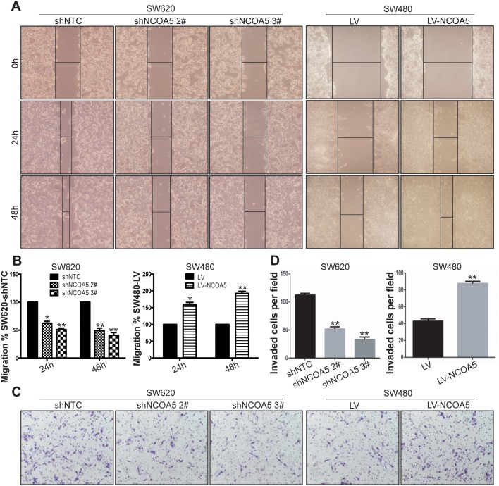
Figure 5. Knockdown of NCOA5 suppresses CRC cell migration and invasion, whereas forced expression of NCOA5 enhances these processes.
(A and B) Wound healing assay. The SW620-shNCOA5 2#, SW620-shNCOA5 3# and SW620-shNTC; SW480-LV-NCOA5 and SW480-LV CRC cells were scratched in the monolayer with a pipette tip. Wound closures were photographed at 0, 24 and 48 hours after wounding. The representative figures of wound healing assay were shown (A). The relative migratory ability of control group was presented (B). SW620-shNCOA5 2# compared with SW620-shNTC group: *, P<0.05 at hour 24, and **, P<0.01 at hour 48; SW620-shNCOA5 3# compared with SW620-shNTC group: **, P<0.01 at hour 24 and 48; SW480-LV-NCOA5 compared with SW480-LV group: *, P<0.05 at hour 24, and **, P<0.01 at hour 48, two-way repeated measures ANOVA, n=6 replicates per condition. (C and D) Transwell invasion assay. The representative figures of Transwell invasion assay were shown (C). The number of invaded tumor cells was counted (D). SW620-shNCOA5 2# and SW620-shNCOA5 3# compared with SW620-shNTC group: **, P<0.01, one-way repeated measures ANOVA; SW480-LV-NCOA5 compared with SW480-LV group: **, P<0.01, Student t test, n=3 replicates per condition, n=10 observations per replicate. Data shown were representative of four independent experiments.

