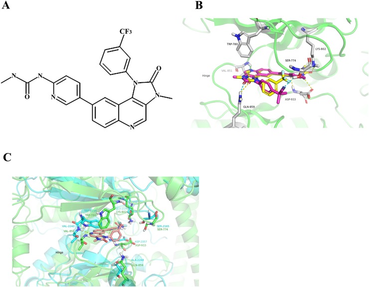
Figure 1.
(A) Chemical structure of SHR8443. (B) The binding modes of BEZ235 and SHR8443 with PI3Kα. The protein was represented as a ribbon diagram (green); SHR8443 (yellow) and BEZ235 (magenta), as well as residues that interacted with these compounds, were shown in stick form. Hydrogen bonds were shown as dashed lines (SHR8443, yellow; BEZ235, cyan) between heavy atoms. (C) The binding mode of SHR8443 within mTOR. SHR8443 was represented by wheat-colored sticks; mTOR and PI3Kα were shown as cyan and green ribbon diagrams, respectively. The key residues of mTOR and PI3kα were shown as sticks. Hydrogen bonds were shown as dashed lines (yellow) between heavy atoms.

