
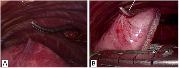
Figure 3. Resection of pGGO under VATS.
(A) A right lateral approach VATS view of the right upper lung. (B) The wedge resection directly guided by Hookwire with no obviously visible subpleural lesion.
Official websites use .gov
A
.gov website belongs to an official
government organization in the United States.
Secure .gov websites use HTTPS
A lock (
) or https:// means you've safely
connected to the .gov website. Share sensitive
information only on official, secure websites.
(A) A right lateral approach VATS view of the right upper lung. (B) The wedge resection directly guided by Hookwire with no obviously visible subpleural lesion.