
Figure 2.
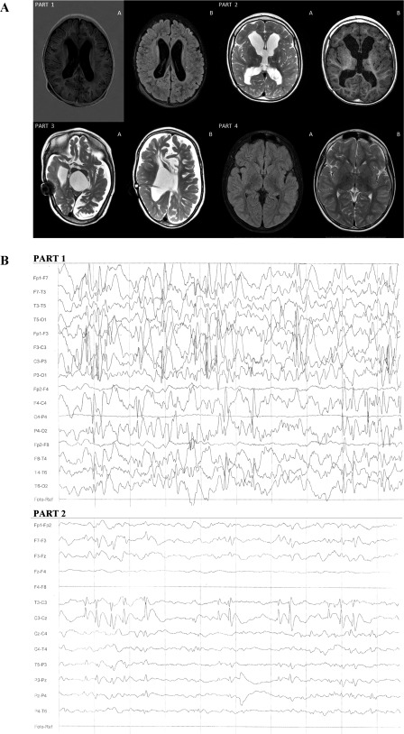
Magnetic resonance imaging (MRI) analysis and electroencephalography (EEG) evaluation. (A): MRI analysis. Part 1A: Patient 1 MRI. T1W turbo inversion recovery magnitude (TIRM) signs of active hydrocephalus; dilatation of lateral ventricles appeared with slightly increased intracranial pressure. Part 1B: Signs of blood‐brain barrier damage resulted in irregular density of the brain cortex, slightly increased intracranial pressure. Part 2A: Patient 2 MRI. T1W, hydrocephalus—winded lateral ventricles and third ventricle, without signs of increased intracranial pressure; blood‐brain barrier damage resulted in irregular density of the brain cortex. Part 2B: T2W, hydrocephalus without signs of activity, without increased intracranial pressure; signs of the destruction of the nuclei basales. Part 3A: T1W diffuse hypoxic destruction of white and grey matter and nuclei basales—post inflammatory vast areas of periventricular white matter malacia; vast areas of white matter and cortex atrophy. Part 3B: T1W—signs of active hydrocephalus with wide lateral, third, and fourth ventricles and increased intracranial pressure after implantation of ventriculoperoneal shunt; vast areas of white matter and cortex atrophy. Part 4A: T2 trim dark fluid‐destruction of nuclei basales; no signs of increased intracranial pressure. Part 4B: T2 TSE—diffuse hyperintensive angiogenic and demyelination regions in white matter, especially in frontotemporal lobes, and minimal focal changes in nuclei basales; no signs of increased intracranial pressure. (B): EEG evaluation. Part 1: Patient 2 EEG taken before treatment—hypersynchronous sleep EEG activity with groups and series of slow theta waves, single and groups of sharp waves, groups of spike‐and‐slow‐wave complexes (1–2 seconds duration), delta waves discharge located on right sight; the spike‐and‐slow‐wave complexes had higher amplitude, even 200 µV, with tendency to generalization; photostimulation and hyperventilation did not affect EEG activity. Part 2: Patient 2 EEG taken after last round of bone marrow mesenchymal stem cells showed reduction focal discharges—sharp waves of spike‐and‐slow‐wave complexes percentage reduction with curtailment of tendency to generalization, smaller percentage of delta waves discharge located on right sight; photostimulation and hyperventilation did not affect EEG activity.