
Figure 1.
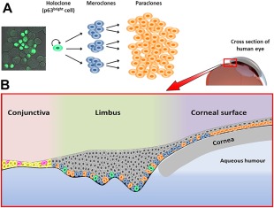
The role of clonogenic keratinocytes in generation and renewal of the corneal epithelium. (A): The holoclone differentiation process from highly proliferative self‐renewing holoclones to transiently amplifying cells (meroclones and paraclones). A confocal microscopy image of holoclone stem cells is on the left showing high expression of ΔNp63α, an isoform of the p63 transcription factor. Due to this characteristic, these cells are also referred to as p63bright cells. (B): Stem cells from (A) in their ocular context. Holoclones, meroclones, and paraclones are found in the basal layer of the limbus with holoclones having the least abundance (10%–15%). The basal layer of the cornea is populated by meroclones and paraclones at the periphery, and only paraclones in the central cornea. All suprabasal layers of the limbus and corneal surface contain terminally differentiated cells which have no capacity for self‐renewal or proliferation (shown in gray). The conjunctival surface is composed of epithelial cells (yellow) and a low proportion of goblet cells (magenta) that are interspersed throughout.