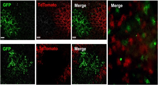
Figure 3.

Three‐dimensional analysis by two‐photon microscopy reveals discrete green or red lung patches after transplantation of a 1:1 mixture of GFP and TdTomato lung cells. A 1:1 mixture of GFP and TdTomato adult lung cells was transplanted into recipient mice following conditioning. Mice were sacrificed and examined by two‐photon microscopy 8 weeks after transfer. Discrete green or red patches were found in the host lung. The absence of yellow patches strongly indicates the clonal origin of each patch. Left: Upper and lower panels illustrate two typical fields at high magnification (extended focus image showing entire scan depth of chimeric lung; each z step = 1 µm, merge of 30–80 planes; scale bar = 50 µm). Right: Image of a larger field at low magnification (scale bar = 200 µm). Abbreviation: GFP, green fluorescent protein.
