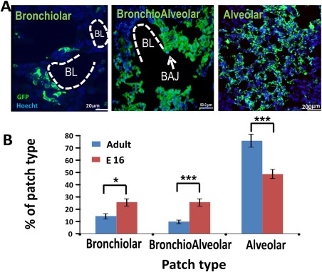
Figure 4.

Different patch types found in the host lung after transplantation. Host mice were sacrificed 8 weeks post‐transplantation and their lungs were evaluated by immuno‐histology. (A): Three major types of patches were identified according to their anatomical position, namely, alveolar (scale bar = 20 µm), bronchiolar (scale bar = 50 µm), and bronchoalveolar (scale bar = 200 µm). BL and BAJ areas are bolded in the figures. Note that the GFP positive, donor derived cells, are present in both alveolar and bronchiolar regions in the host lung. (B): Percentage of patch types out of the total number of patches. These results (n = 6 mice, eight fields were counted for each mouse) were compared to results found after transplantation of 1E + 6 E16 fetal lung cells (n = 3 mice, 10 fields were counted for each mouse). Errors bars represent mean ± SD. t test was used to analyze statistical significance between the E16 and the adult groups in each section. *, p < .05; **, p < .01; ***, p < .001. Abbreviations: BAJ, broncho‐alveolar junctions; BL, bronchiolar lumen; GFP, green fluorescent protein.
