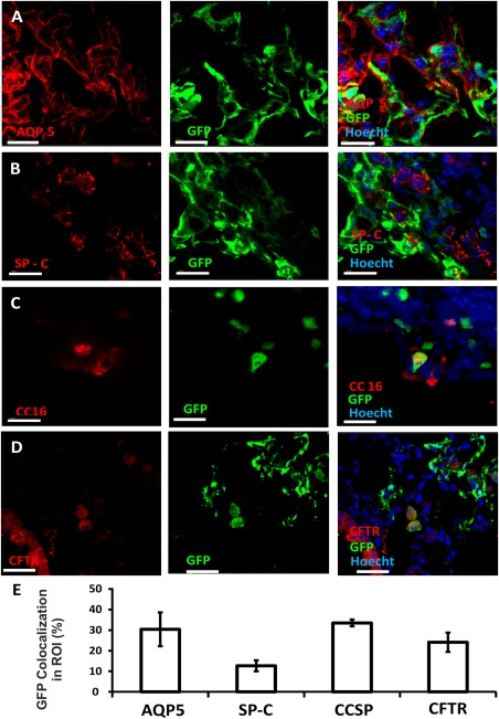
Figure 6.

Integration of donor‐derived cells in the epithelial lung compartment. Immunohistological analysis was used to characterize the composition of the epithelial compartment within donor‐derived GFP+ green patches. Imaris software was used to determine fluorescence colocalization. GFP+ areas were designated as the areas of interest, and the colocalization channel identified pixels that included both red and green colors. ATI cells were stained with Aquaporin type 5 (A), ATII cells with surfactant protein C (B), club cells with CCSP (CC16) (C), and CFTR+ cells with anti‐CFTR (D). In each figure, the red channel represents the lineage marker, the green channel marks donor‐derived GFP+ cells, and the blue channel marks nuclear staining. Scale bar = 40 µm. (E): Summary of % ROI (average plus SD) colocalized 8 weeks after transplantation of 8E + 6 adult GFP+ lung cells (based on five different areas obtained from two transplanted mice). Abbreviations: GFP, green fluorescent protein; ROI, region of interest.
