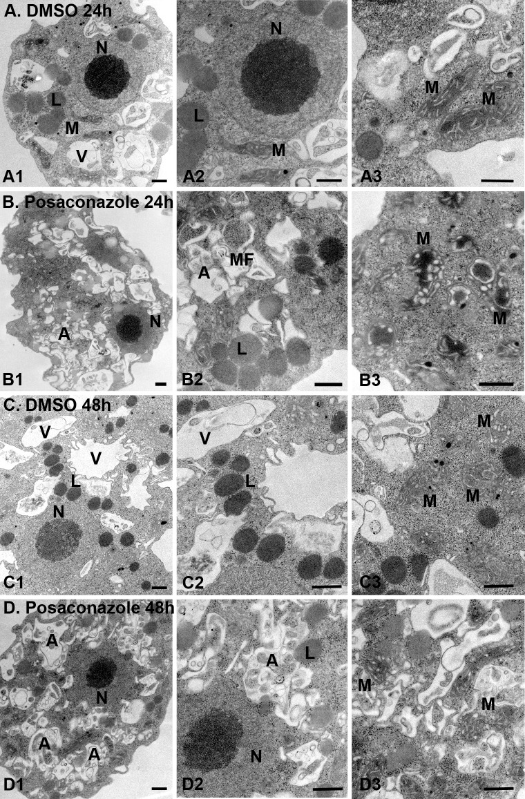
Fig 5. Ultrastructural analysis of posaconazole-treated N. fowleri by transmission electron microscopy (TEM).
0.1% DMSO-treated controls (A: A1-A3, 24 h; and C: C1-C3, 48 h, show several food vacuoles (V), mitochondria (M), nucleus (N) and lipid droplets (L). Exposure to 0.2 μM posaconazole (B: B1-B3, 24 h; D: D1-D3, 48 h) led to mitochondrial swelling, accumulation of atypical lipid droplets, alteration of nuclear membrane and appearance of autophagic vacuoles (A) engulfing organelle debris and myelin figures (MF). Bar = 500 nm.

