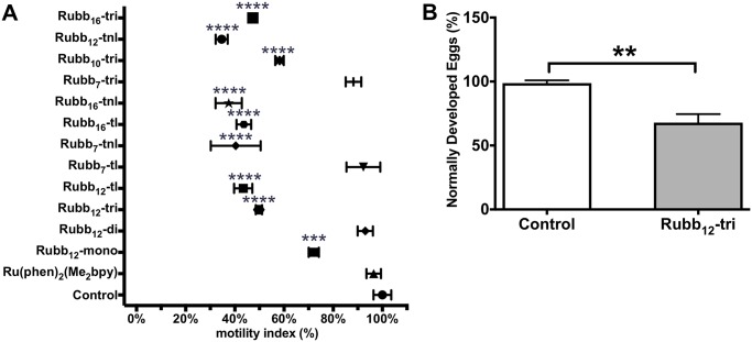
Fig 4. Inhibition of S. mansoni egg hatching and effect on egg development by ruthenium complexes.
(A) Graph representing the percentage of S. mansoni eggs hatched (motility index) in the presence of various ruthenium complexes (50 μM) as determined by the x-WORM motility assay. Data represents the average of triplicate experiments ± SE. (B) Triplicate sets of five pairs of adult S. mansoni worms were cultured in Basch media with or without 5 μM Rubb12-tri for 72 h. The eggs released into the media were counted and those that were misshapen or immature were scored as “abnormally developed”. Graph shows the difference in percentage of normally developed eggs between treated and control groups and data represents the average of triplicate experiments ± SE. Differences in egg hatching were measured by ANOVA and differences in egg development by t test. *P ≤ 0.05, **P ≤ 0.01, ***P ≤ 0.001,****P ≤ 0.0001.

