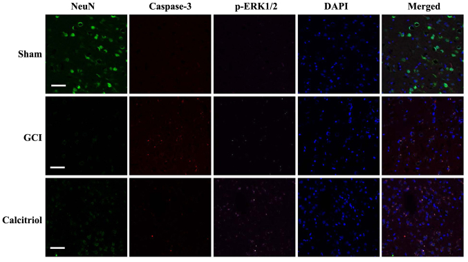
Figure 9.
Fluorescence staining for NeuN, caspase-3 and p-ERK1/2 in rat cortex, and the merged images. Immunofluorescence staining was performed at 3 days in Sham, GCI and calcitriol groups (n=5 for each group; scale bar, 50 μm; green, NeuN; red, caspase-3; purple, p-ERK1/2; blue, DAPI). GCI, global cerebral ischemia; p-ERK, phosphorylated extracellular signal-regulated kinase.

