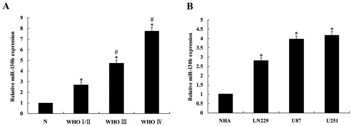
Figure 1.
Expression levels of miR-130b in glioma tissues and cell lines. (A) miR-130b was detected in 30 glioma tissues and 5 non-neoplastic brain specimens by reverse transcription-quantitative polymerase chain reaction. miR-130b expression was significantly higher in the glioma tissues compared with the non-neoplastic brain specimens, and the expression in the high level glioma groups is higher than in the low level group. *P<0.05 vs. non-neoplastic brain specimens; #P<0.05 vs. WHO I/II group. (B) miR-130b expression was detected in LN229, U87 and U251 glioma cell lines and primary NHAs. qPCR showed that miR-130b was upregulated in all of the glioma cell lines compared with the NHA cells. *P<0.05 vs. NHA cells. NHA, normal human astrocytes.

