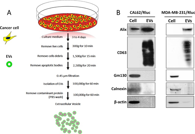
Figure 1. Isolation and characterization of EVs.
(A) A flow chart for the EV purification procedure based on ultracentrifugation. (B) Western blotting analysis of EVs. ALIX and CD63, EV marker proteins were detected by using anti-ALIX (97 kDa) and anti-CD63 (53 kDa) specific antibodies, respectively; GM-130 and Calnexin, EV negative marker proteins were detected by using anti-GM-130 (112 kDa) and anti-calnexin (90 kDa) specific antibodies, respectively; β-actin was used as a loading control.

