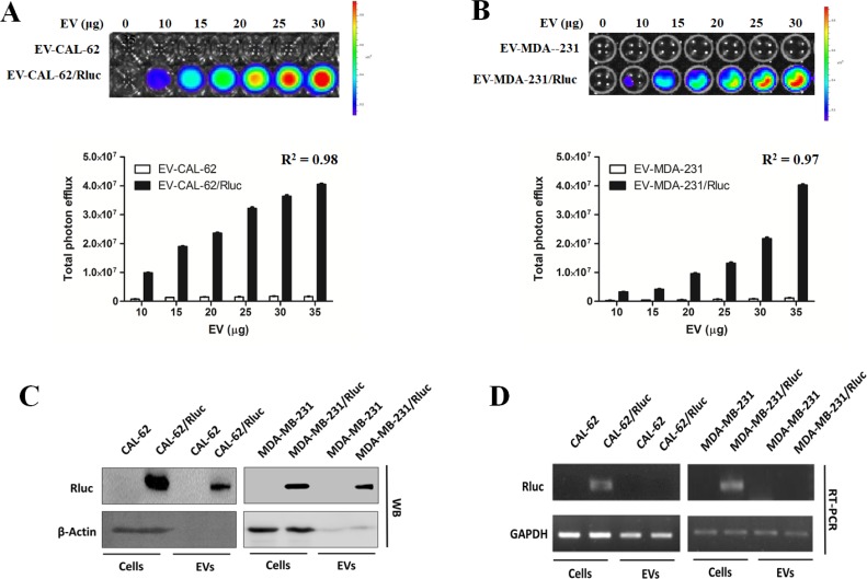
Figure 3. EV-CAL-62/Rluc showed EV-specific Rluc activity in vitro.
(A, B) Representative bioluminescent imaging of an in vitro luciferase assay in EVs from CAL-62, CAL-62/Rluc cells, MDA-MB-231 and MDA-MB-231/Rluc. Quantitative in vitro luciferase assay in EV's data are expressed as mean ± SD. (C) Western blot analysis of the Rluc protein in cells and EVs from CAL-62, CAL-62/Rluc & MDA-MB-231 and MDA-MB-231/Rluc cells, detected by means of Rluc-specific antibodies. β-Actin served as loading control. (D) RT-PCR analysis of Rluc mRNA in cells and EVs from CAL-62, CAL-62/Rluc & MDA-MB-231 and MDA-MB-231/Rluc cells. GAPDH was used as a loading control.

