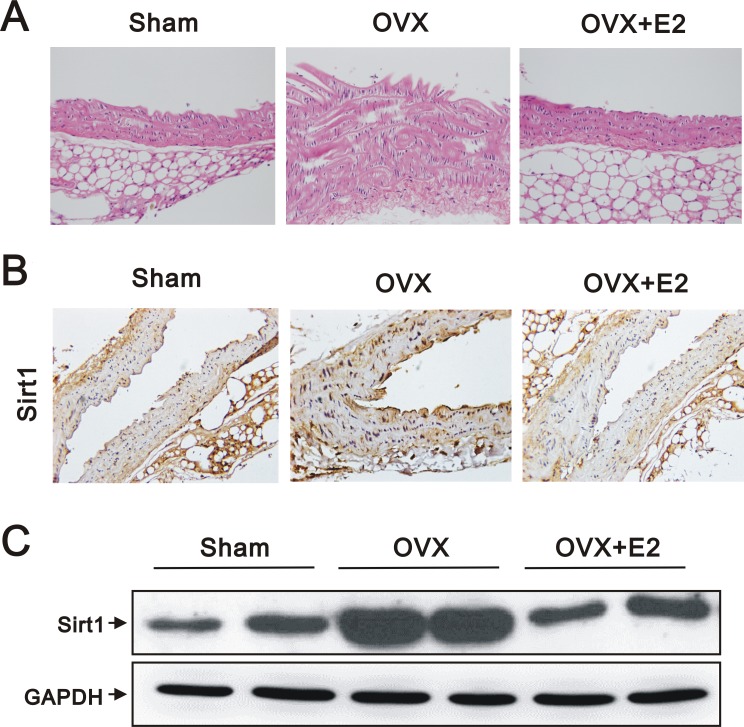
Figure 1. Histology, IHC, and western blot analyses of aortas from sham, OVX, and OVX+E2 mice.
(A) Representative images of hematoxylin and eosin stained, paraffin-embedded aortas (original magnification, 400×), (B) representative IHC staining for SIRT1 expression (original magnification, 400×), and (C) western blots using anti-SIRT1 and anti-GAPDH antibodies of aortic tissues from sham, OVX, and OVX+E2 mice.

