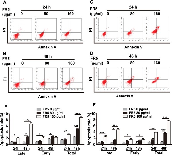
Figure 3. Apoptosis-induced effect of FR5 in Bel7402 and HepG2 cells.

Bel7402 and HepG2 cells were planted into 6-well plates at 2 × 10⁵ cells/well, treated with FR5 at 0 (Ctrl), 80 and 160 μg/ml for 24 and 48 h, stained with Annexin V-FITC/PI, and detected by flow cytometry. (A, B) The apoptosis rate of HepG2 cells at 24 h and 48h of FR5 incubation, respectively. (C, D) The apoptosis rate of Bel7402 cells at 24h and 48 h of FR5 incubation, respectively. (E, F) Histogram shows the difference of HepG2 and Bel7402 apoptotic cells (%) between time and doses. Results were presented as mean ± SD, and the error bars represent the SD of three independent experiments. *p<0.05; **p<0.01; ***p<0.001 vs control group.
