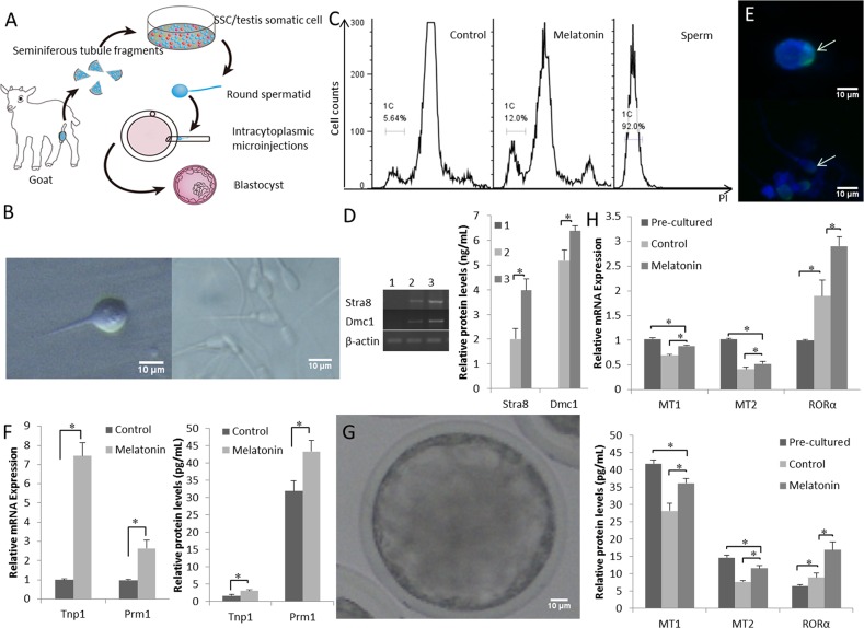
Figure 2. Functional goat haploid spermatozoa were obtained from the in vitro testis cell culture.
(A) A schematic illustration of the culture system used in the present study. (B) Representative micrographs of a spermatid with a single flagellum isolated from the in vitro culture (left) and adult goat sperm used as a control (right). (C) DNA contents of the suspended cultured cells were examined by flow cytometry. “Control” is the group that was induced to differentiate with basic medium. “Melatonin” represents cells cultured in basic medium supplemented with melatonin. Adult sperm cells were used as a positive control. 1C marks the haploid peaks. (D) The expression of Stra8 and Dmc1 was assessed in suspension cells. 1: cells from the original passage; 2: cells passaged after differentiation; 3: after induction with 10-7 M melatonin, suspension cells were passaged after differentiation. (E) Haploid cells expressed the mature sperm protein acrosin (green); nuclei of the cells were stained with DAPI (upper panel), and adult goat sperm were used as a control (lower panel). (F) Expression patterns of post-meiotic genes (Tnp1 and Prm1). (G) Reconstructed embryos developed to the blastocyst stage. (H) The expression of the MT1, MT2, and RORα was detected pre-culture and post-culture; differentiated cells and cells cultured without melatonin were used as controls. Data are expressed as means ± SEM. * P < 0.05.

