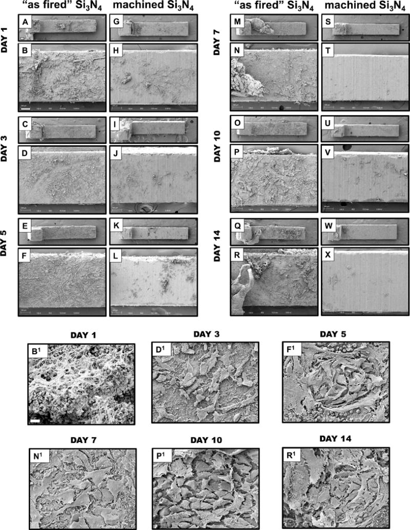
Figure 4. Differential host cell responses to sterile “as fired” vs. machined Si3N4 implants in bone over time.

Sterile L-shaped “as fired” and machined Si3N4 implants (n=5) were surgically implanted into the tibiae of 6-week-old, female Balb/c mice, removed on the indicated day post-op, and processed for SEM (A-X). Representative low power images of the L-shaped implants are shown at × 30 (bar = 200μm), and higher power images of regions containing host material are shown at ×125 (bar =100μm). Of note is the highly reactive “as fired” Si3N4 implant surface, which displayed a progressive transformation towards osseous integration over time. Day 1 white and red blood cells (B, B1×1000); Day 3 rounded/oval cells (mesenchymal stem cells (MSCs)) (D, D1×1000); Day 5 rounded to polygonal cells (differentiation MSCs) (F, F1×1000); Day 7 Large polygonal mesenchymal cells (osteoblasts) (N, N1 ×1000); Day 10 & Day 14 osteoblastic cells organizing to form a dense coating (P, P1 ×1000 & R, R1 ×1000, respectively). In contrast, machined Si3N4 implant surfaces were largely non-reactive with the host, and did not display any remarkable changes over time.
