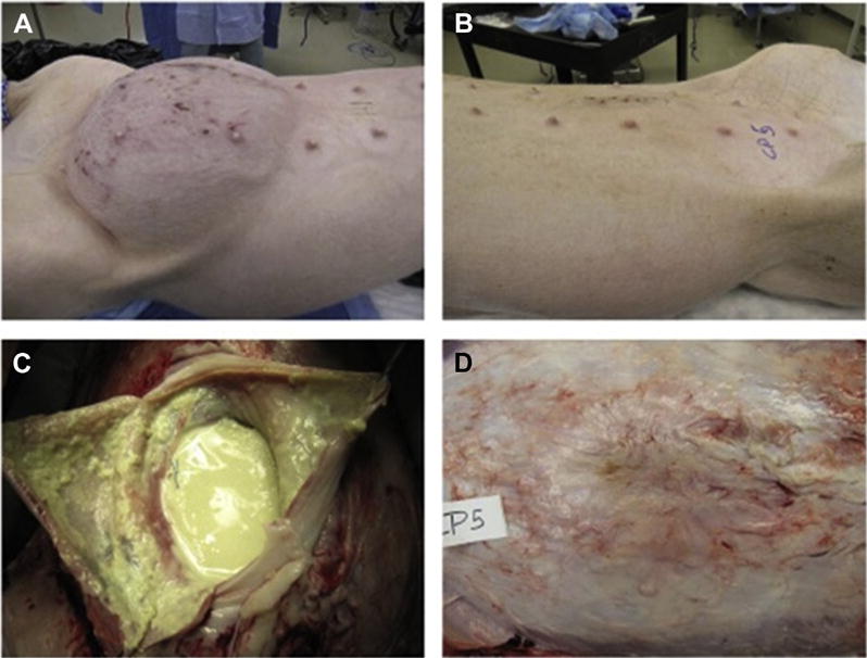
Fig. 1.

Images taken at the study end point (30 d). These images are representative of animals in the infected/untreated mesh condition (A and B) and in the antibiotic delivery mesh condition (C and D). Control animals with no infection appeared similar to (C and D) and are not shown. Infected/untreated animals showed extensive signs of infection as well as inflammatory/immunological response (B) in addition to significant abdominal distension (A). Animals receiving antibiotic delivery meshes showed excellent hernia repair (C) and minimal inflammation and scarring on the abdominal flap (D). (Color version of figure is available online.)
