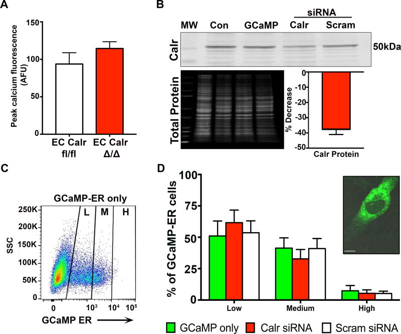
Figure 5. Calreticulin knockdown in endothelial cells does not affect the level of endoplasmic reticulum calcium.
A, Third order mesenteric arteries from EC Calr fl/fl and EC Calr Δ/Δ mice were loaded with Fluo-4AM. Arteries were incubated in 0 mM extracellular calcium, along with cyclopiazonic acid (20µM) to inhibit SERCA refilling of ER calcium stores. This provides an indirect measurement of ER calcium stores (EC Calr fl/fl n=3; EC Calr Δ/Δ n=3, compared using unpaired student's t-test). B, Representative western blot of primary human aortic EC, which were transfected with GCaMP-ER plasmid to specifically detect ER calcium fluorescence. Then EC were transfected with either calreticulin siRNA or scrambled siRNA. Knockdown was assessed via western blot for calreticulin. Values were normalized to total protein for each lane.and compared to scrambled siRNA. Control EC were untransfected. MW=molecular weight ladder. C, Representative flow cytometry plot for EC transfected with GCaMP-ER to visualize ER calcium, divided into low (L), medium (M) and high (H) fluorescent signal. SSC=side scatter. D, Quantification of GCaMP-ER signal for EC co-transfected with Calr or scrambled siRNA. Inset: Representative confocal image of an EC with GCaMP-ER signal (green, visualized with GFP laser) indicating calcium within the ER. (GCaMP-ER only n=3, Calr siRNA n=3, Scram siRNA n=3, compared using one-way ANOVA. No comparisons were made between Low/Med/High expression.) Scale bar=10µm.

