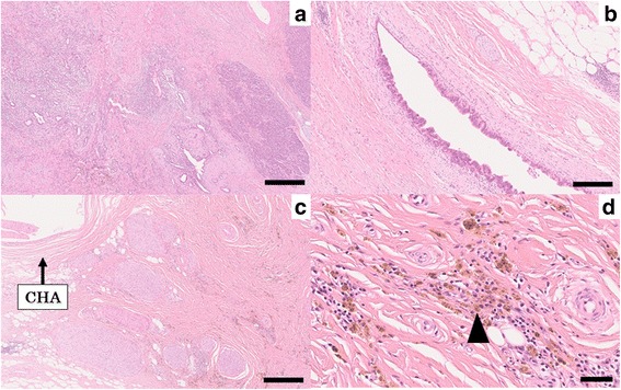
Fig. 5.

Representative histological photographs of resected specimen (hematoxylin and eosin staining). a The viable adenocarcinoma cells invaded into pancreatic tissue with the formation of various ductal structures (×50, bar; 500 μm). b The tumor cells extended to the main pancreatic ducts (×100, bar; 250 μm). c The tissue around the CHA was replaced with fibrous tissue (×50, bar; 500 μm). d The fibrous tissue included many histiocytes containing hemosiderin (arrowhead) (×400, bar; 50 μm)
