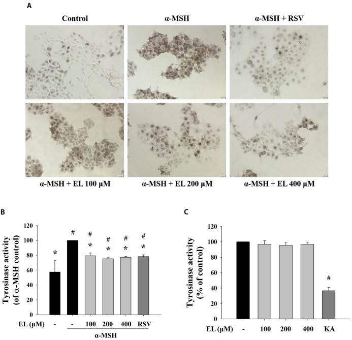
Fig. 2. Effects of ethyl linoleate on tyrosinase activity in B16F10 cells.
The B16F10 cells were exposed to α-melanocyte-stimulating hormone (α-MSH, 500 nM) in the presence of ethyl linoleate (EL) for 48 h. (A) In situ intracellular tyrosinase activity determined by L-DOPA staining. Resveratrol (RSV, 20 µM) was used as a positive control. Images were captured under identical conditions using bright field microscopy. Bar=20 µm. (B) Intracellular tyrosinase activity was determined using lysates obtained from B16F10 cells treated with EL or RSV. (C) The direct effect of ethyl linoleate on tyrosinase activity was measured with mushroom tyrosinase. Kojic acid (KA, 400 µM) was used as a positive control. The data are expressed as the means±SD. *p<0.01, compared with the α-MSH control; #p<0.01, compared with the vehicle control.

