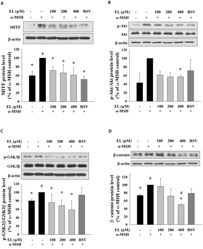
Fig. 4. Effects of ethyl linoleate on expression of MITF through Akt/GSK3β/β-catenin signal in B16F10 cells.
The B16F10 cells were exposed to α-melanocyte-stimulating hormone (α-MSH, 500 nM) in the presence of ethyl linoleate (EL) or resveratrol (RSV, 20 µM) for 4 h. The expression levels of protein including Akt, p-Akt, GSK3β, p-GSK3β, β-catenin, and β-actin were detected by using western blot. Band intensity compared to the α-MSH control was determined by ImageJ software. (A) MITF, (B) Akt and p-Akt, (C) GSK3β and p-GSK3β, and (D) β-catenin. The data are expressed as the means±SD. *p<0.05, compared with the α-MSH control; #p<0.05, compared with the vehicle control.

