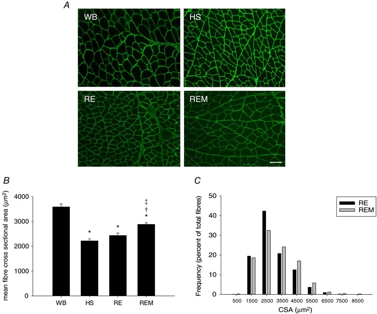
Figure 2. Massage is associated with enhanced recovery of cross sectional area (CSA) during reambulation.

A, representative images of gastrocnemius muscle cross sections of WB, HS, RE and REM rats immunostained for dystrophin (green). Scale bar = 50 μm. B and C, quantification of muscle fibre CSA of gastrocnemius from WB (n = 8), HS (n = 7), RE (n = 8) and REM (n = 8) rats (B), and frequency distribution of CSA in RE (n = 8) and REM (n = 8) rats only (C). Values are means ± SEM. One‐way ANOVA followed by Tukey's post hoc test was used to determine statistical significance: *different from WB, †different from HS, ‡difference from RE, P < 0.05.
