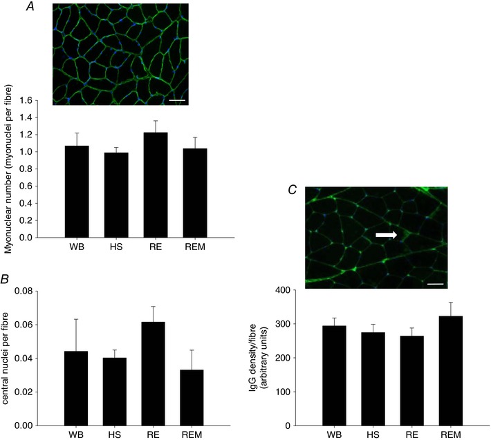
Figure 7. No increase in myonuclear number or overt damage to muscle fibres with massage.

Representative image of dystrophin (green) and DAPI (blue) staining with myonuclear number (A) and central nuclei (B) quantified, and a representative image of IgG staining (green) with the white arrow pointing to intramuscular IgG, which is quantified (C) in gastrocnemius muscle of WB (n = 8), HS (n = 7), RE (n = 8) and REM (n = 8) rats. Scale bar is 50 μm. Values are means ± SEM. One‐way ANOVA was used to determine statistical significance (P < 0.05).
