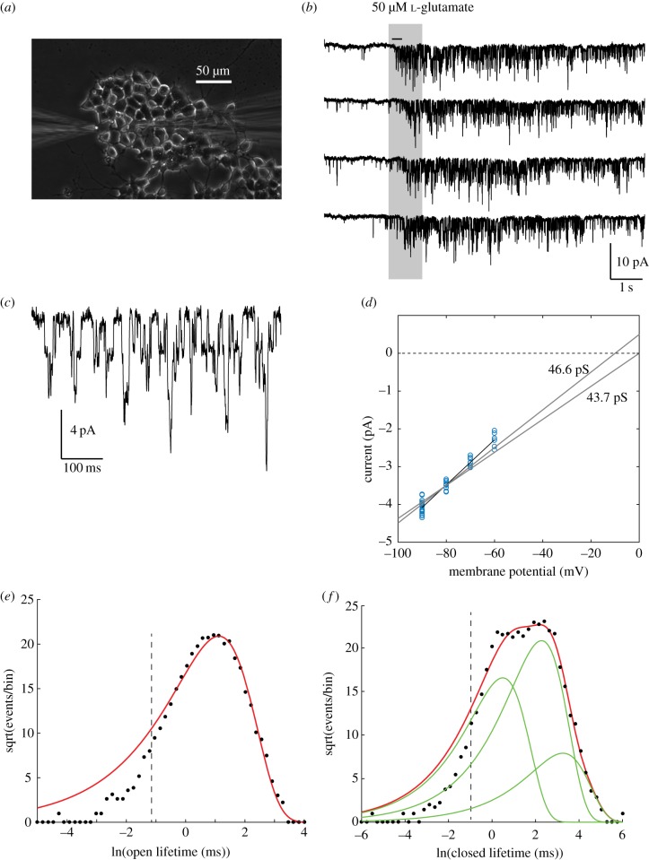
Figure 1.
Functional NMDARs in PanNET cells. (a) Puff application of glutamate. Puffer pipette at left, patch pipette at right. (b) Whole-cell patch recording, 500 Hz Gaussian digital low-pass filter, −80 mV. (c) Expanded segment of top trace in (b) (indicated by horizontal bar) showing single-channel opening and closing transitions. (d) Current–voltage plot of channel openings. Each point is data from one cell at one holding potential (n = 21 cells). Best-fit slope conductance (γ) for reversal potential (Erev) of 0 mV was 46.6 pS; and for Erev = −5 mV, γ = 43.7 pS. (e,f) Sigworth-Sine plots [13] of channel lifetime distributions in one cell. (e) Open-state durations fitted to a single exponential distribution (6129 events, bin size = 0.18, missed events cut-off < 0.331 ms indicated by dashed vertical line). τ = 3.06 ms. (f) Closed-state durations, fitted to a sum of three exponential components (8183 events, bin size = 0.24, a1 = 0.318, τ1 = 1.63 ms, a2 = 0.595, τ2 = 9.75 ms, a3 = 0.09, τ3 = 26.0 ms.

