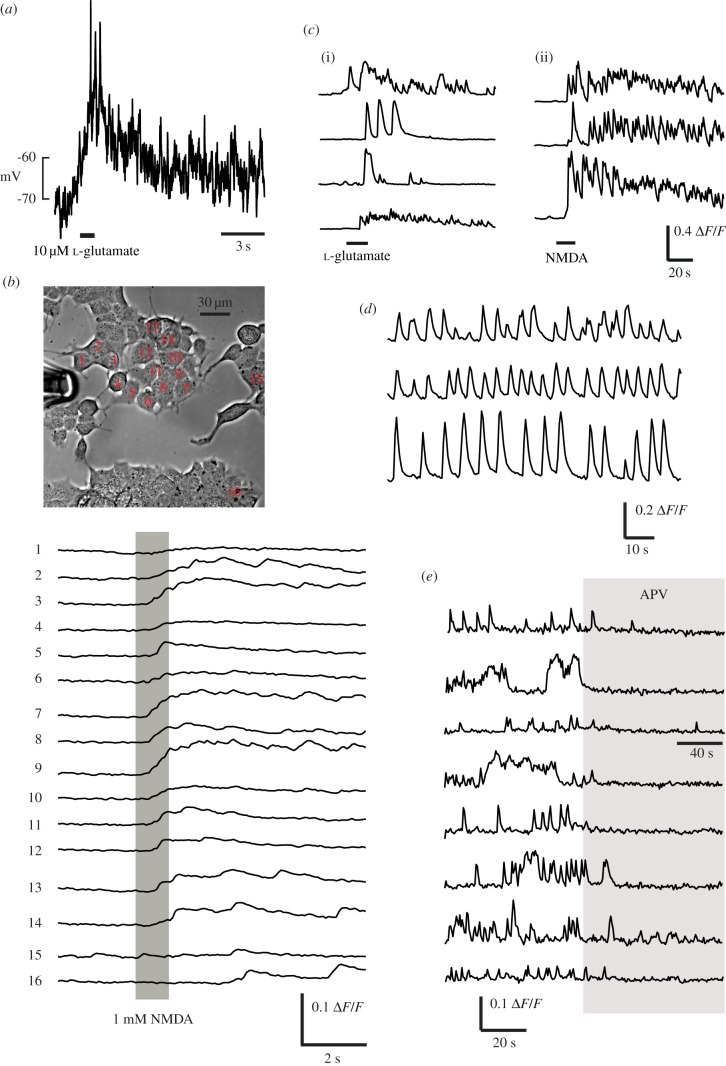
Figure 2.
NMDARs can stimulate membrane depolarization and calcium influx. (a) whole-cell current-clamp recording with high [K+] solution in the pipette. A 1s pulse of l-glutamate containing Ringer solution (500 µM) was applied through a perfusion pipette at the time indicated, causing a large, maintained depolarization accompanied by action potentials. (b) After loading with Oregon Green BAPTA-1AM, fluorescence was recorded while puff-applying 1 mM NMDA dissolved in the Ringer solution. Synchronous elevations in intracellular calcium are observed in cells across the field which are exposed to the specific agonist. 90 of 173 cells analysed (52%) showed detectable responses (average ΔF/F 6.44% in responding cells) to pressure ejection of glutamate or NMDA. (c) Example calcium responses in several cells to (i) 20 µM l-glutamate or (ii) 1 mM NMDA. (d) Three simultaneously-recorded cells showing spontaneous calcium transients, at 35°C in 1 mM Mg2+ extracellular solution. (e) Examples of APV-induced silencing of spontaneous activity in cells from eight different culture dishes. Perfusion with 1 mM APV occurs during the period indicated by the grey bar.

