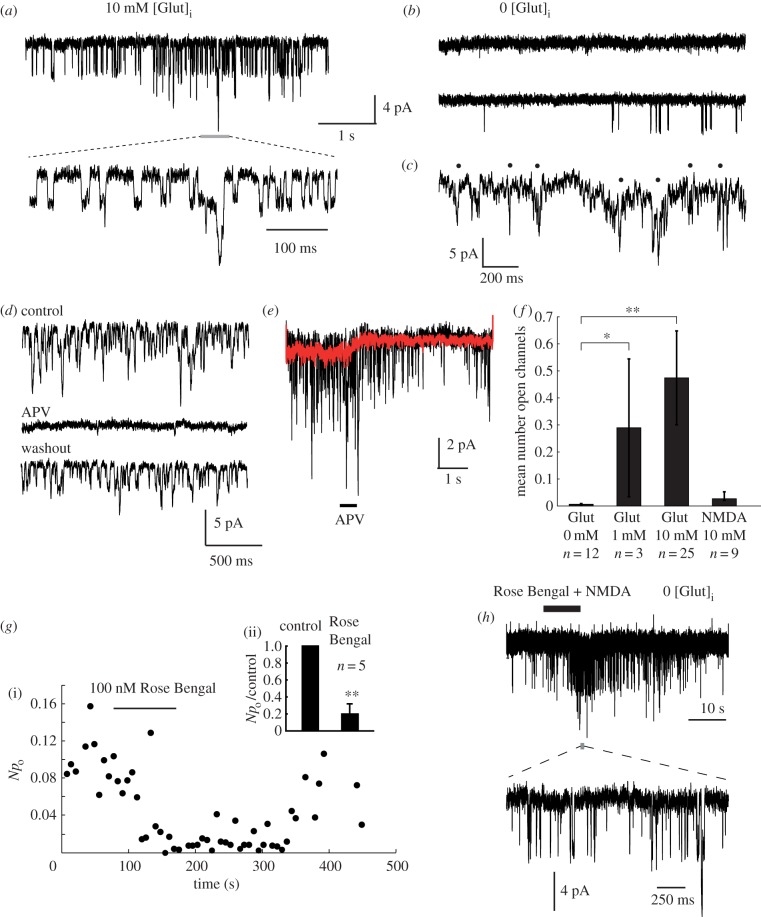
Figure 3.
Autocrine activation of NMDARs. (a) A constant NMDAR channel activation is observed at −90 mV using pipettes filled with a caesium gluconate based solution containing 10 mM l-glutamate (autocrine condition). A segment of channel activity at higher time resolution is shown below, as indicated. (b) Very infrequent or no NMDAR activation is seen when glutamate is omitted from the patch pipette (paracrine condition). (c) Nystatin-perforated patch recordings show poorly-resolved 4–5 pA openings (indicated by black spots). (d) Channel activity is completely and reversibly blocked by perfusion with 1 mM APV. (e) Puff-perfusion of APV solution for 500 ms causes a rapid block in channel openings. Black trace: individual trial. Red trace: ensemble average of 20 successive trials. (f) Summary of the channel activity produced by intracellular agonists. The ordinate is the average number of open channels measured over a 5 s period at −90 mV. Significance: control versus 1 mM Glut, *p < 0.0484; control versus 10 mM Glut, **p < 0.000026; control versus 10 mM NMDA, p > 0.458; Wilcoxon rank sum test. (g) Sensitivity of autocrine NMDAR activation to a VGLUT inhibitor: (i) Reversible block of autocrine activity by 100 nM Rose Bengal. Npo is the product of channel number and open probability, equivalent to the mean number of simultaneously-open channels. (ii) Rose Bengal (100 nM) block is significant, **p < 0.0079, Wilcoxon rank test, n = 5 cells. (h) Rose Bengal (100 nM) does not block responses to exogenously applied NMDA (1 mM) in cells recorded with zero glutamate in the pipette (paracrine condition). A short segment of channel activity is shown at high time resolution below, as indicated.

