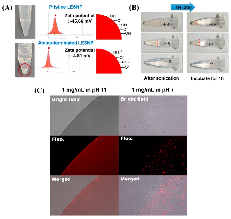
Figure 6.
(A) comparison of the dispersion behaviors of the amine-terminated LESNP under different pH conditions. The upper panel shows the image of a pristine LESNP and its zeta-potential analysis. The lower panel shows the image of an amine-terminated LESNP and its zeta-potential analysis; (B) images of the amine-terminated LESNP in three different types of buffer solutions at different pHs (pH 3, 7, and 11). The aggregation of particles under the three different conditions was observed for one hour; (C) the result of a fluorescence microscopy analysis of the amine-terminated LESNP in two different buffer solutions of pH 7 and pH 11, respectively.

