
Figure 6.
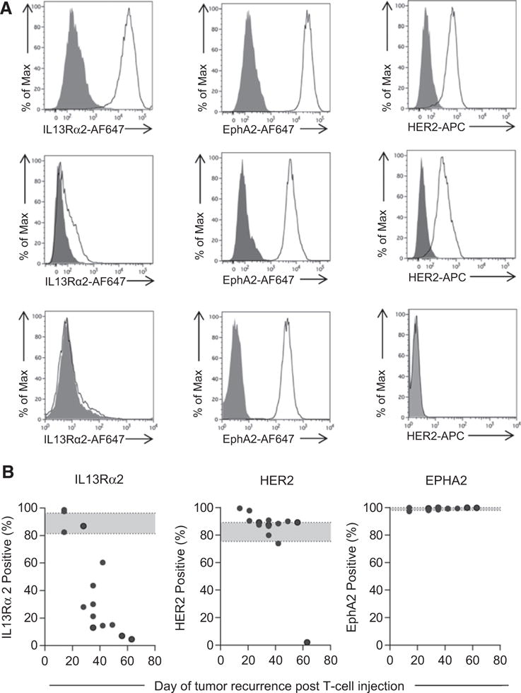
Late-recurring U373 gliomas did not express IL13Rα2. Cells isolated from recurrent tumors were analyzed for IL13Rα2, EphA2, and HER2 expression using primary goat anti-IL13Rα2 (AF146, R&D Systems), mouse anti-EphA2 (MAB3035, R&D Systems), and HER2-APC (340554, BD Biosciences) followed by secondary (except for HER2) rabbit anti-goat or goat anti-mouse IgG Alexa647 (Life Technologies). A, Representative FACS plots. Top row, IL13Rα2-positive recurrent tumors; middle row, IL13Rα2 low-expressing recurrent tumors; bottom row, IL13Rα2-negative recurrent tumors; gray, isotype control; and white, antigen-specific antibody. B, Summary data of GBM-associated antigen (IL13Rα2, HER2, EphA2) expression; each dot represents a recurring tumor (n = 13; one independent experiment per one recurrence, total 13).