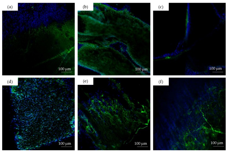
Figure 8.
CLSM images (×20) of: (a) brain; (b) intestine; (c) skin; (d) blood vessels; (e) cornea; and (f) gonad cells on hydrogel surfaces after organotypic culture. Cell nuclei (blue) were labeled using Hoechst 33,342 (blue), and the fibrin network (green) was stained using an FITC conjugated to anti-fibrinogen antibody.

