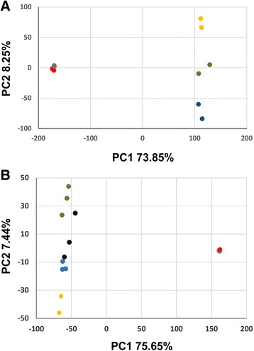
Fig. 3.

Embryogenic and callus cells resemble octant stage embryos and the peripheral domain of the SAM. Embryogenic cells and callus cells are shown in red and gray respectively. Each dot represents an experimental replicate. a PCA comparing callus and embryogenic cells to early stages of embryo development. Seeds carrying 1–2 cell embryos, 8 cell embryos (octant), and 32 cell embryos (globular stage) samples are shown in yellow, green, and blue, respectively. b PCA comparing callus and embryogenic cells to various sub-domains of the SAM. Stem cell niche marked by CLV3 expression, SAM without CLV3 expressing domain, SAM organizing center marked by WUS expression, and SAM peripheral zone marked by FILAMENTOUS FLOWER (FIL) expression samples are highlighted in green, black, yellow and blue, respectively
