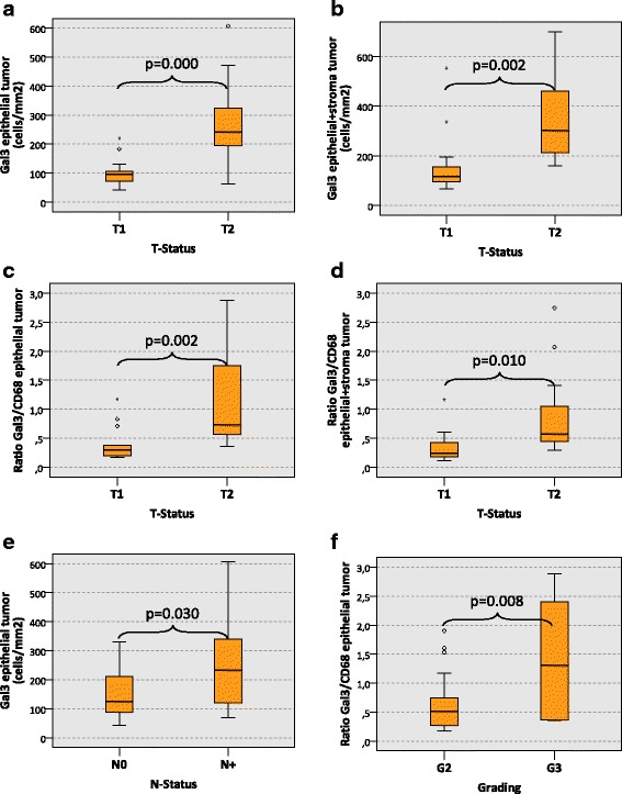
Fig. 3.

Galectin 3 expression in tumor resection specimens of oral squamous cell carcinomas depending on histomorphologic parameters. The a and b show the expression of Galectin 3 (Gal3) (cells/mm2 specimen area) in the epithelial compartment (a) and in the whole analyzed specimen area (epithelial + stroma) (b) of oscc tumor resection specimens depending on the T-status (T1 vs. T2). A significantly increased count of Gal3-positive cells can be found in the epithelial compartment and in the whole analyzed specimen area of T2 oscc tumor resection specimens. The c and d show the ratio between the Gal3 cell count and the CD68 cell count (Gal3/CD68 ratio) in the epithelial compartment (c) and in the whole analyzed specimen area (epithelial + stroma) (d) of oscc tumor resection specimens depending on the T-status (T1 vs. T2). A significantly increased Gal3/CD68 ratio can be found in the epithelial compartment and in the whole analyzed specimen area of T2 oscc tumor resection specimens. e shows the expression of Gal3 (cells/mm2 specimen area) in the epithelial compartment of oscc tumor resection specimens depending on the N-status (N0 vs. N+). A significantly increased count of Gal3-positive cells can be found in N+ oscc tumor resection specimens. f shows the Gal3/CD68 ratio in the epithelial compartment of oscc tumor resection specimens depending on the grading (G2 vs. G3). A significantly increased Gal3/CD68 ratio is detected in the epithelial compartment of G3 oscc tumor resection specimens. P-values generated by the ANOVA test are indicated in all boxplots
