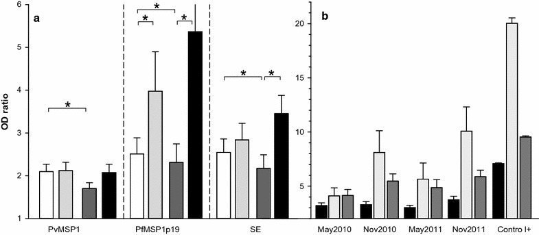
Fig. 2.

Levels of IgG responses to P. falciparum and P. vivax as function of time of follow-up. Longitudinal fluctuation of antibody levels is plotted in part a as histogram + SE for each Ag in May 2010 (white), November 2010 (light grey), May 2011 (dark grey), November 2011 (black). Asterisks indicate significant different level of Ab (P < 0.05, Wilcoxon signed rank test). In panel b, antibody levels for all positive responders (individuals with ODratio > 2) to PvMSP1 (black), PfMSP1p19 (light grey), SE (dark grey) are shown as histogram plots + SE for each set of samples from May 2010 to November 2011. IgG levels of positive controls are also shown for comparison
