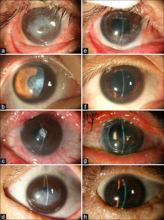
Figure 1.

Representative pre- and post-operative photos of patients receiving DALK. (a) A 39-year-old male patient suffered from chemical burn OD. The corneal was reconstructed by conjunctivolimbal autograft with residual stromal opacity, and the vision was CF/30 cm. (e) The graft remained clear 5 years after DALK, and the BCVA reached 20/60. (b) A 18-year-old male suffered from HSV stromal keratitis OD, BCVA was 20/250. (f) Microperforation of DM was experienced during surgery, but the graft remained clear 5 years after DALK. The endothelial cell count was 1947/mm2, and BCVA was 20/25. (c) A 31-year-old female was a case of SJS with corneal scarring, neovascularization, and extreme thinning. Central corneal perforation OS was sealed with Histoacryl glue, and the vision was HM/20 cm. (g) Four years after DALK, the graft remained clear. Limited by preexisting advanced glaucoma, the BCVA was CF/60 cm. (d) An 11-year-old boy received meningioma excision with resulting neurotrophic and exposure keratitis OS. The vision was CF/10 cm. (h) The patient received DALK combined with permanent tarsorrhaphy. Four years after DALK, the graft remained clear, and the BCVA improved to 20/25 (DALK = Deep anterior lamellar keratoplasty, OD = Right eye, OS = Left eye, CF = Counting fingers, HM = Hand motion, HSV = Herpetic simplex virus, BCVA = Best-corrected visual acuity, SJS = Stevens–Johnson syndrome, DM = Descemet's membrane)
