
Figure 1. Structures of Apo and Agonist-bound TRPML3.
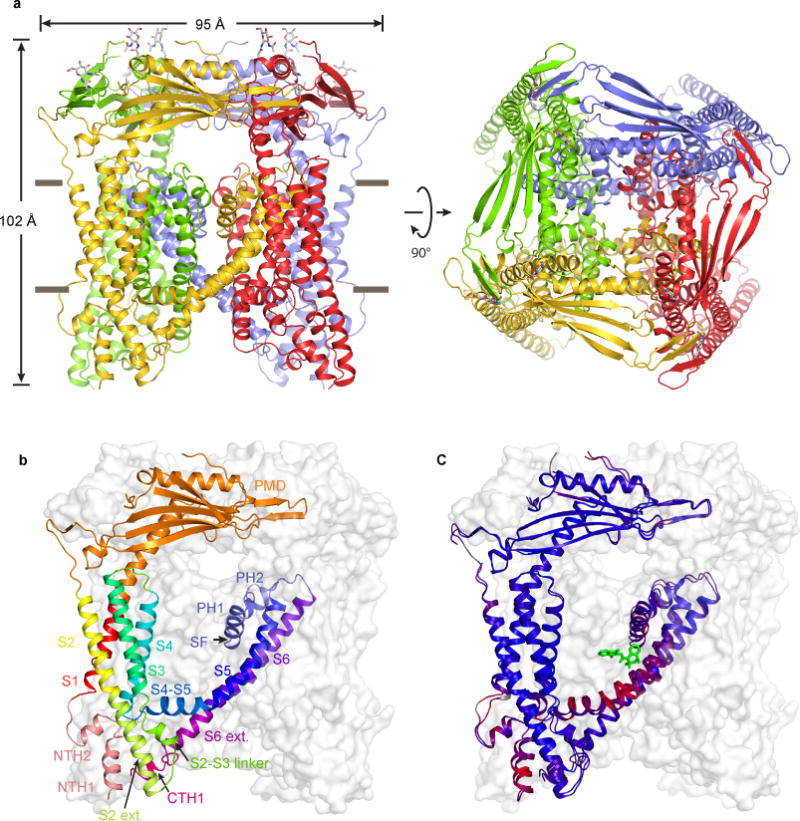
(a) ML-SA1-bound structure, viewed parallel to the membrane (left) and from the luminal/extracellular side of the membrane (right). (b) Structure of a protomer, viewed parallel to the membrane. Different regions are illustrated in different colors. The other three subunits are shown in grey surface representations. (c) Superposition of the apo and ML-SA1-bound structures. Blue and red indicate similarity and divergence, respectively.