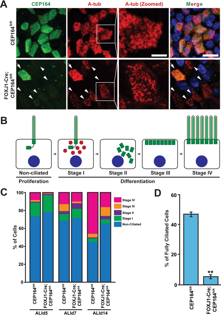
Fig 4. Ablation of CEP164 leads to defective airway multiciliogenesis.
(A) ALId14 MTECs were stained for CEP164 (green) and A-tub (red). Nuclei were stained with DAPI (blue). Arrowheads denote CEP164-KO multiciliated cells with sparse, stubby cilia. Zoomed views of cilia are shown for the squared areas. Arrows indicate a multiciliated cell with CEP164 expression that escaped Cre-mediated recombination in FOXJ1-Cre;CEP164fl/fl MTEC cultures. Scale bars, 10 μm and 5 μm for zoomed images. (B) Schematic model depicting the stages of multiciliated cell differentiation. See text for details. (C) Quantification of multiciliated cells at different stages (I-IV) of ciliogenesis. MTECs from CEP164fl/fl and FOXJ1-Cre;CEP164fl/fl mice were fixed at ALId5, d7, and d14 and immunostained for A-tub. n>225 total cells per ALI day from each of three independent MTEC preparations per genotype. (D) Quantification of fully ciliated cells. MTECs from CEP164fl/fl and FOXJ1-Cre;CEP164fl/fl mice were fixed at ALId14 and immunostained for A-tub. The percentages were calculated by dividing the number of fully ciliated cells with abundant cilia by total cell number. n>250 total cells from each of three independent MTEC preparations per genotype. Error bars represent ±SEM. **, p<0.01.

