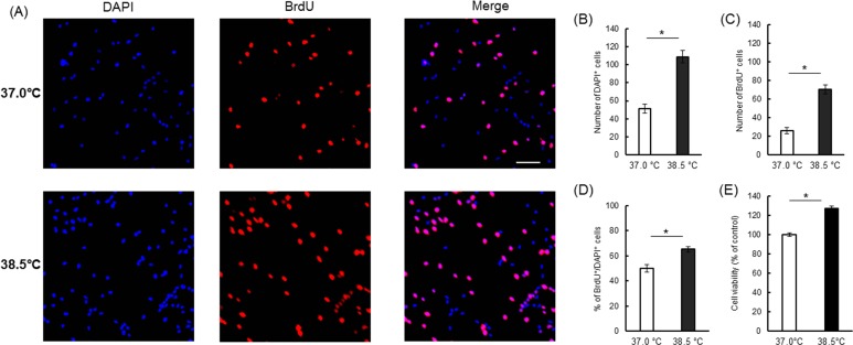
Fig 2. Proliferative effects of mild heat exposure on NSCs/NPCs in a monolayer culture.
(A) Representative images of BrdU-labelled cells (red) in control (37.0°C) and heat-exposed (38.5°C) conditions. Nuclei are counterstained by DAPI (blue). Scale bar (white) is 50 μm. Bar diagrams showing the number of total cells (B), number of BrdU+ cells (C) and percentage of BrdU+/DAPI+ cells (D) at 37.0°C and 38.5°C on day 3 of monolayer attached NSC/NPC culture in PM. Results are expressed as mean ± SEM of three independent experiments. *P < 0.05 vs. control condition. (E) On day 4 of monolayer culture in PM, cell viability was measured by MTS assay. Data are expressed as percentages of the control condition (37.0°C). Results are mean ± SEM of three independent experiments. *P < 0.05 vs. control condition.

