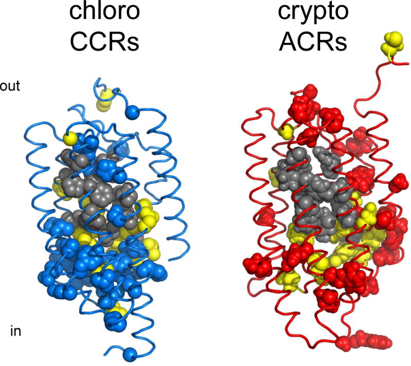
Figure 6.

Structural comparison of chlorophyte CCRs and ACRs. Gray, residues of the retinal binding pocket of BR conserved in each of the two types of channelrhodopsins; yellow, residues conserved in both CCRs and ACRs; blue, residues conserved only in CCRs; red, residues conserved only in ACRs. The residue conservation pattern is shown using the C1C2 crystal structure (3ug9; left) and a GtACR1 homology model built on the 3ug9 template (right).
