. 2017 Dec 14;6:e27215. doi: 10.7554/eLife.27215

Figure 1. Cell-intrinsic enhanced ability of Ly-6C- CD4 TN cells to commit into iTreg cells.

(A–E) Flow-cytometry sorted Ly-6C- and Ly-6C+ CD4 TN cells from C57BL/6 Foxp3-GFP mice were stained with CTv (Ly-6C-) or CTv and CTfr (Ly-6C+) and stimulated separately or together for 4 days with coated αCD3 and αCD28 Abs (4 µg/mL), in the presence of graded doses of TGFβ1. (A) Diagram illustrating the experimental protocol. (B) Representative CTv/CTfr dot-plots for gated CD4+ cells recovered after 4 days of culture. Ly-6C- and Ly-6C+ CD4 TN cells were either cultured separately (top left and right panels, respectively) or together (bottom panel) (C) Representative Foxp3/CD4 contour-plots and proportions of Foxp3+ cells for gated CD4+ cells are shown at a dose of 0.25 ng/mL TGFβ1. (D) Proportions of Foxp3+ cells among CD4+ cells are shown as a function of TGFβ1 concentration. Mean ± s.e.m of four independent experiments are shown. (E) Concentrations of TGFβ1 needed to obtain 50% of the maximal percentages of iTreg-cell polarization (EC50) were calculated for each CD4 TN cell subset in separated or mixed cultures. Each dot represents an independent experiment. Significance of differences were assessed using a two-tailed paired Student’s t-test. Values of p<0.05 were considered as statistically significant (**p<0.01; ns, not significant).

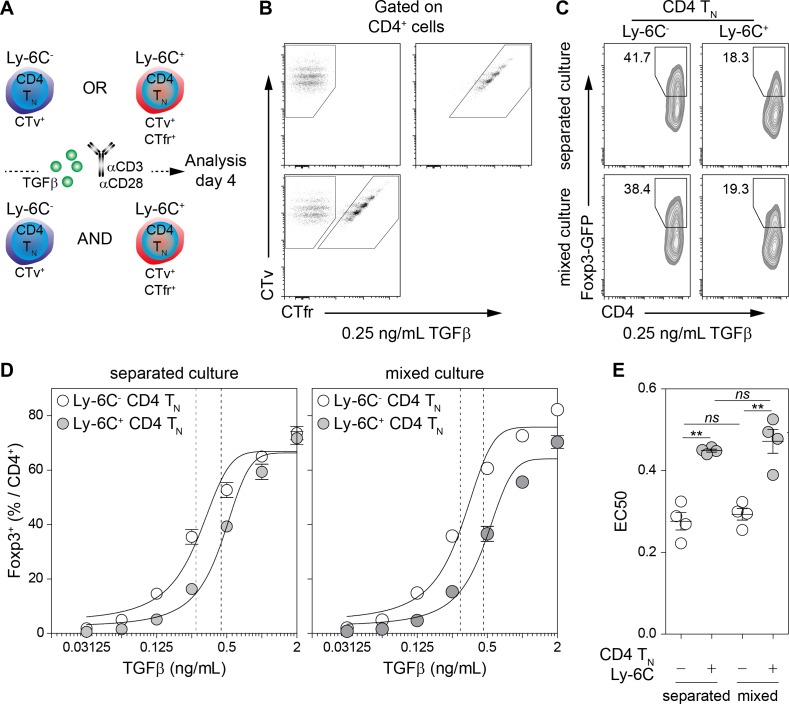
Figure 1.

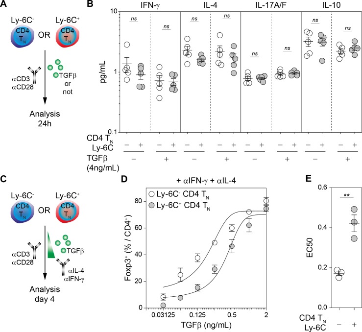
Figure 1—figure supplement 1. Cytokine-independent enhanced ability of Ly-6C- CD4 TN cells to commit into iTreg cells.

Figure 1—figure supplement 1.

(A–B) Flow-cytometry sorted Ly-6C- and Ly-6C+ CD4 TN cells from C57BL/6 Foxp3-GFP mice were stimulated separately with coated αCD3 and αCD28 Abs (4 µg/mL), in the presence of TGFβ1 (4 ng/mL) or not. 24 hr later, culture supernatants were collected and analyzed for the presence of interferon-gamma (IFN-γ) and interleukins (IL) −4, −17A/F and −10. (A) Diagram illustrating the experimental protocol. (B) IFN-γ, IL-4, IL-17A/F and IL-10 concentrations in the culture supernatants of both Ly-6C- and Ly-6C+ CD4 TN cells after 24 hr culture are shown. (C–E) Flow-cytometry sorted Ly-6C- and Ly-6C+ CD4 TN cells from C57BL/6 mice were stimulated separately with coated αCD3 and αCD28 Abs (4 µg/mL), in the presence of αIFN-γ and αIL-4 blocking antibodies and graded doses of TGFβ1. (C) Diagram illustrating the experimental protocol. (D) Proportions of Foxp3+ cells among CD4+ cells are shown as a function of TGFβ1 concentration. Mean ± s.e.m of two independent experiments are shown. (E) Concentrations of TGFβ1 needed to obtain 50% of the maximal percentages of iTreg-cell polarization (EC50) were calculated for each CD4 TN-cell subset in separated or mixed cultures. Each dot represents an independent experiment. Significance of differences were assessed using a two-tailed paired Student’s t-test. Values of p<0.05 were considered as statistically significant (**p<0.01; ns, not significant).