
Fig. 1.
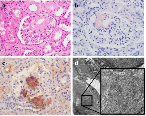
Histological features of the non-cancerous renal tissue. a Deposition of amorphous eosinophilic material was observed at vascular poles and mesangial regions (hematoxylin-eosin stain). The samples were both Congo red- (b) and amyloid A-positive (c). d Randomly dispersed, rigid, nonbranching fibrils were evident on electron microscopy. The width of the fibrils was 8 to 15 nm, and their length was 300 to 1000 nm (original magnification, a, b, c, ×400)