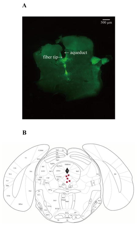
Figure 5. Placement of fiberoptic cannula tips in the DR.
A, an example of coronal brain slice, showing the location of a fiberoptic cannula tip in the DR of a transgenic DBA/1 mouse. B, distribution of representative locations of fiberoptic cannula tips (squares) in the DR, according to the mouse atlas of Paxinos and Franklin (Paxinos and Franklin, 2013).

