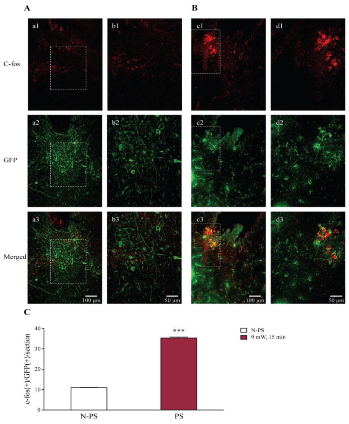
Figure 6. Photostimulation increases c-fos expression in 5-HT neurons in the DR of transgenic DBA/1 mice.
A, neuronal immunostaining of c-fos (a1, b1), GFP (a2, b2) and co-localization c-fos and GFP (a3, b3) in a transgenic DBA/1 mouse that was implanted with fiberoptic cannula, but no photostimulation was applied (n = 3 mice). B, immunostaining of c-fos (c1, d1), GFP (c2, d2) and co-localization c-fos and GFP (c3, d3) in an implanted transgenic DBA/1 mouse with photostimulation at 9 mW for 15 min (n = 3 mice). C, quantification of c-fos(+)/GFP(+) cells in implanted transgenic DBA/1 mice without photostimulation (blank bar) and those in implanted transgenic mice with photostimulation (red bar). N-PS, no photostimulation; PS, photostimulation. Confocal image magnifications: a1–a3 and c1–c3, 20x; b1–b3 and d1–d3, 40x. See figure 2 for photostimulation parameters.
*** p < 0.001: Significantly different from the number of c-fos(+)/GFP(+) cells per section in implanted transgenic DBA/1 mice with no photostimulation.

