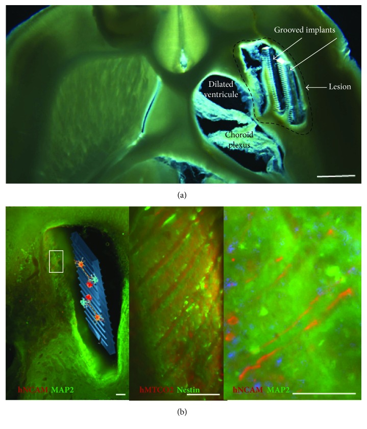
Figure 2.
Representative horizontal brain section of the lesioned area of rats with implants alone (a) (scale bar: 1 mm) and neuro-implants (b) under brightfield illumination. The newly generated tissue was mostly located around the PDMS implants. (b) Human neural stem cells were identified by a specific human marker hNCAM or hMTCO2, in combination with a marker (in green) of immature (nestin) and mature (MAP2) neurons. Low magnification is provided on the left and higher magnifications on the right (scale bars: 100 μm). Grafted cell neurites were aligned along the grooves of the implant.

