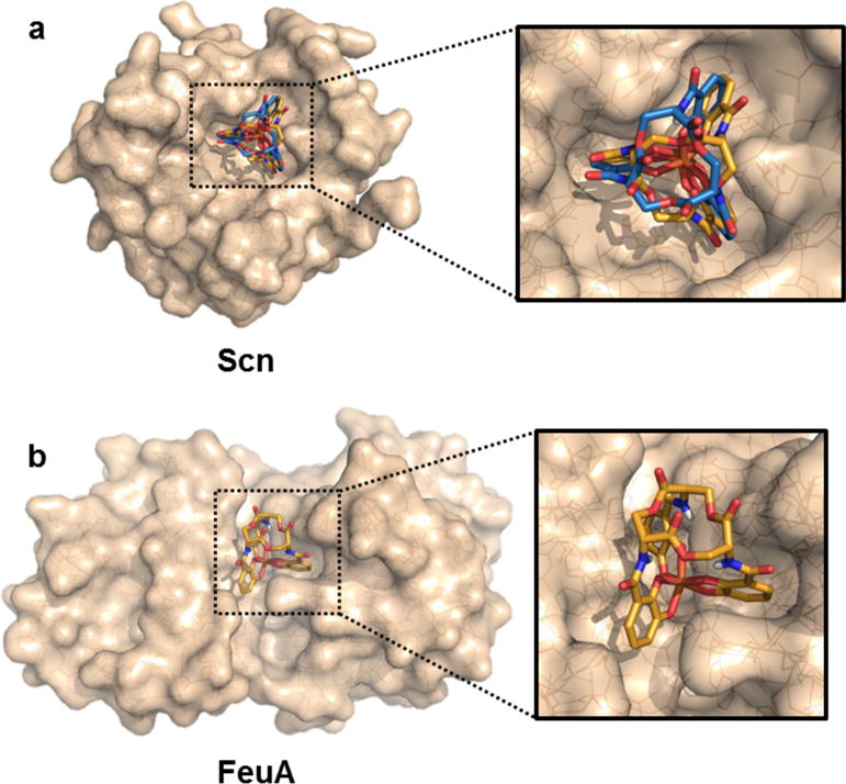
Figure 4.

Protein docking studies with [Fe(D-ent)]3− shown as sticks with orange carbon atoms, [Fe(ent)]3− shown as sticks with blue carbon atoms, and proteins shown as translucent wheat surfaces encasing lines. (a) The crystallographically-determined structure of [Fe(D-ent)]3− docked into FeuA (PDB: 2XUZ). [Fe(ent)]3− could not be successfully docked into the binding site. (b) The crystallographically-determined structures of [Fe(D-ent)]3− and [Fe(D-ent)]3− docked into Scn (PBD: 1L6M).
