Fig. 4.
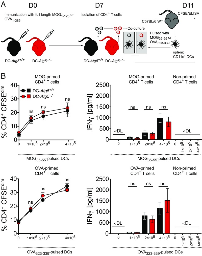
Loss of ATG5 in DCs does not impair priming of antigen-specific CD4+ T cells. (A) Experimental setup. (B) Quantification of DC-Atg5−/−– and DC-Atg5+/+–derived CD4+ T cell proliferation via carboxyfluorescein succinimidyl ester (CFSE) dilution in the presence of increasing amounts of wild type-derived peptide-pulsed DCs (Upper and Lower Left). CD4+ T cell response (IFNγ secretion) is unchanged upon coculture with peptide-pulsed DCs (Upper and Lower Right). Pooled data of two independent experiments are shown. Statistical analysis: Mean ± SEM is depicted. Unpaired two-tailed Student t test was applied. ns, not significant: P > 0.05. DL, detection limit.

