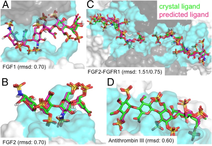
Fig. 2.
Comparison of predicted binding sites for heparin (magenta) to the X-ray crystal (green) ligand positions for the four validation systems. The rmsd between predicted and crystal structures of ligand heavy atoms (A) FGF1 (rmsd of 0.70 Å), (B) FGF2 (rmsd of 0.70 Å), (C) FGF2–FGFR1 (rmsds of 1.51 Å, 0.75 Å), and (D) α-antithrombin III (rmsd of 0.60 Å).

