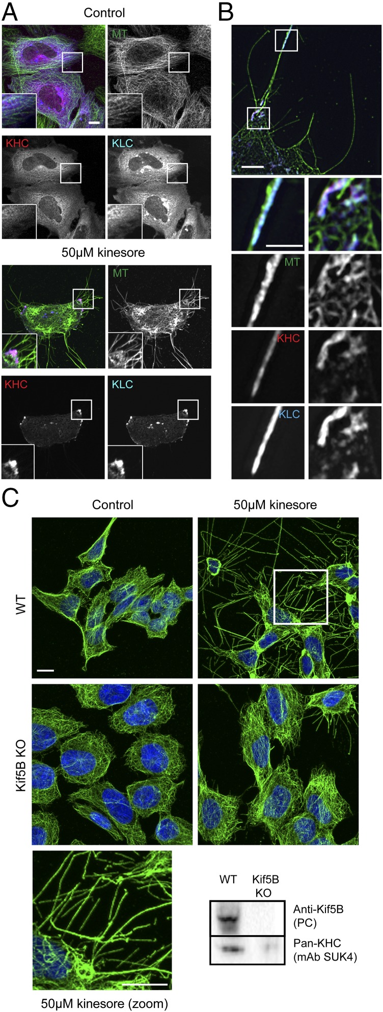
Fig. 3.
Kinesore-induced remodeling of the microtubule network is dependent on kinesin-1. (A) Representative maximum-intensity projection confocal immunofluorescence images showing HeLa cells transfected with mCit-KHC (pseudocolored red) and HA-KLC (pseudocolored blue) treated with 50 μM kinesore (Right) or vehicle control (Left) (0.1% DMSO) for 1 h. Note the change in localization of kinesin-1 (KHC/KLC) and formation of microtubule-rich projections in kinesore-treated cells. Images are representative of three independent experiments (Scale bar, 10 μm. Enlargement magnification: 2.9×.) (B) Structured illumination images of kinesore-treated cells as in A. (Scale bar in B, 5 μm; 2.5 μm in enlargement.) (C) Representative immunofluorescence images of HAP1 cells (wild-type or Kif5B knockout) treated with 50 μM kinesore or vehicle control (0.1% DMSO) for 1 h and stained for β-tubulin (green) or DNA (Hoechst, blue). Microtubule-rich projections induced by kinesore are strongly suppressed by knockout of Kif5B. (Scale bar, 10 μm.) Western blot shows whole-cell extracts of wild-type or Kif5B knockout HAP1 cells probed with either a Kif5B specific polyclonal antibody or a pan-KHC monoclonal antibody (SU.K.4).

